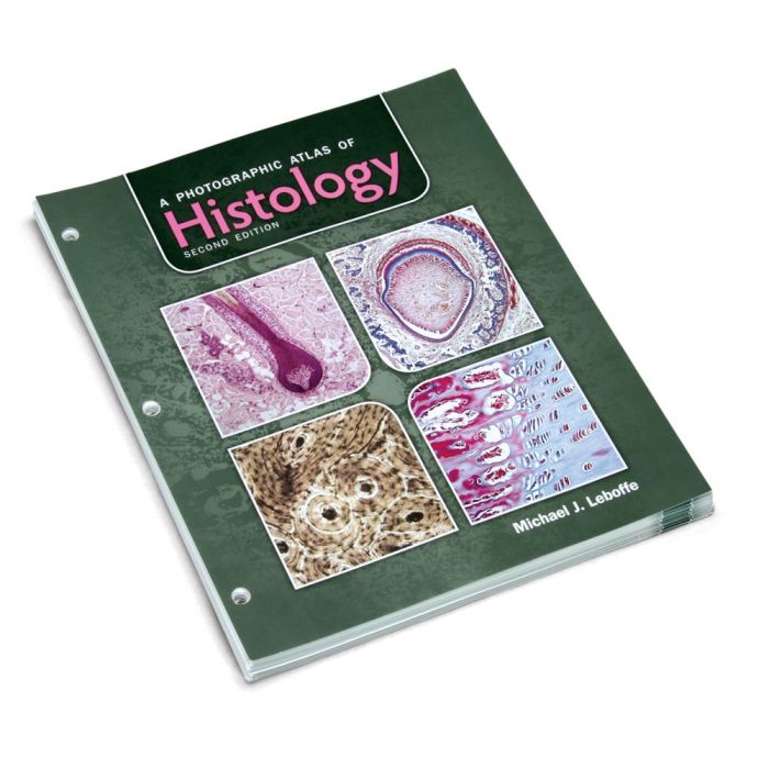
A photographic atlas of histology 2nd edition pdf – Embark on a captivating journey through the microscopic world with “A Photographic Atlas of Histology, 2nd Edition.” This comprehensive resource unveils the intricacies of histological techniques and their profound impact on understanding human biology and disease.
Through stunning high-resolution images and meticulously crafted text, this atlas provides an unparalleled guide to the structural and functional organization of tissues and organs. Prepare to delve into the fascinating realm of histology and unravel the secrets of human health and pathology.
Overview of the Book

The “Photographic Atlas of Histology, 2nd Edition” provides a comprehensive visual guide to the microscopic structure of human tissues and organs. This edition features updated and expanded content, including new images and discussions of emerging histological techniques.
The book is organized into chapters covering the major organ systems, with each chapter providing a detailed overview of the histological features of the tissues and organs within that system.
Histological Techniques and Imaging
Tissue Preparation and Staining
The book covers a wide range of histological techniques, including tissue preparation, staining, and microscopy. These techniques are essential for preserving and visualizing the microscopic structure of tissues.
Imaging Modalities
The book employs various imaging modalities, such as light microscopy, electron microscopy, and immunohistochemistry. Each modality offers unique advantages for visualizing different aspects of tissue structure.
Organization and Structure: A Photographic Atlas Of Histology 2nd Edition Pdf
The book is organized into chapters and sections that cover various tissues and organs. This organization facilitates easy navigation and reference.
Each chapter begins with an introduction that provides an overview of the organ system and its histological features. The chapters are then divided into sections that cover specific tissues and organs within that system.
Image Quality and Presentation

The histological images presented in the book are of exceptional quality, providing clear and detailed views of the microscopic structure of tissues.
The images are well-labeled and accompanied by informative captions that provide additional information about the structures being visualized.
Educational Value
The “Photographic Atlas of Histology, 2nd Edition” is an invaluable educational resource for students and researchers in histology.
The book’s clear and concise text, combined with its high-quality images, makes it an effective tool for teaching and learning about the microscopic structure of human tissues and organs.
Pedagogical Features, A photographic atlas of histology 2nd edition pdf
The book includes several pedagogical features, such as case studies, review questions, and online supplements, that enhance its educational value.
The case studies provide real-world examples of how histological techniques are used in clinical practice.
Comparison with Other Histology Atlases
The “Photographic Atlas of Histology, 2nd Edition” compares favorably with other leading histology atlases.
It offers a comprehensive coverage of histological techniques and imaging modalities, as well as a wide range of high-quality images.
The book’s clear and concise text and its user-friendly organization make it an excellent choice for students and researchers in histology.
Clinical Relevance

The histological images presented in the book have direct clinical relevance.
By understanding the microscopic structure of tissues, healthcare professionals can make accurate diagnoses and provide optimal patient care.
For example, the book’s images can help pathologists identify abnormal cell changes that may indicate disease.
FAQ Guide
What are the key features of “A Photographic Atlas of Histology, 2nd Edition”?
The atlas boasts high-quality histological images, detailed explanations of histological techniques, and a comprehensive organization that facilitates easy navigation.
How can this atlas benefit students and researchers?
The atlas serves as an invaluable educational resource, providing a deep understanding of histological techniques and their applications in biomedical research.
What sets this atlas apart from other histology resources?
Its exceptional image quality, thorough coverage of histological techniques, and emphasis on clinical relevance distinguish this atlas as a standout resource.